Transcriptional dysregulation of autophagy in the muscle of a mouse model of Duchenne muscular dystrophy


Personal take on this article:

Abstract:
This study investigates the transcriptional dysregulation of autophagy in the skeletal muscles of mdx mice, a well-established model for Duchenne muscular dystrophy (DMD). The primary focus is on the roles of FoxO transcription factors (FoxO1 and FoxO3a) and transcription factor EB (TFEB), which are pivotal in regulating autophagy-related genes. The findings reveal significant downregulation of these genes in dystrophin-deficient muscles due to phosphorylation-mediated suppression of FoxOs and TFEB.


Key Findings:


1. Global Downregulation of Autophagy-Related Genes:
Microarray analyses and quantitative PCR revealed a significant reduction in the expression of genes involved in autophagosome formation, autophagosome-lysosome fusion, and mitophagy in the tibialis anterior and soleus muscles of mdx mice compared to wild-type controls.
   

2. FoxO1 and FoxO3a Dysregulation:

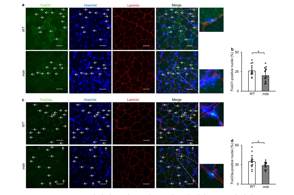
Immunostaining and Western blot analyses showed decreased nuclear localization and increased phosphorylation of FoxO1 (Thr24) and FoxO3a (Thr32), suggesting impaired transcriptional activity due to Akt-mediated phosphorylation.
   

3. TFEB Dysregulation:

The study also demonstrated reduced nuclear localization and increased phosphorylation of TFEB (Ser122) in mdx mice, indicating mTORC1-mediated suppression of TFEB activity.


4. Accumulation of Autophagic Markers:

Elevated levels of LC3-II and p62 proteins were observed, indicating impaired autophagosome degradation and suggesting a disruption in the autophagic flux in dystrophic muscles.


5. Resveratrol as a Potential Therapeutic Agent:

Treatment with resveratrol, an activator of SIRT1, was shown to restore the expression of autophagy-related genes, decrease phosphorylation of TFEB, and ameliorate muscle pathology in mdx mice, highlighting its potential therapeutic benefits.


Mechanistic Insights:
- Akt-mTORC1 Pathway: Enhanced Akt-mTORC1 signaling in dystrophic muscles leads to increased phosphorylation and nuclear export of FoxOs and TFEB, resulting in the downregulation of their target genes involved in autophagy.


- FoxOs and TFEB: Both transcription factors are crucial for the maintenance of autophagic activity. Their suppression contributes to the pathogenesis of DMD by disrupting the autophagic degradation of damaged proteins and organelles, leading to muscle degeneration and atrophy.


Clinical Implications:
The study underscores the importance of targeting the transcriptional regulation of autophagy in developing novel therapeutic strategies for DMD. By correcting the dysregulated autophagy pathway, it may be possible to mitigate muscle degeneration and improve clinical outcomes for DMD patients.


Conclusion:
The research provides compelling evidence that transcriptional dysregulation of autophagy, mediated by phosphorylation-dependent suppression of FoxOs and TFEB, plays a critical role in the pathophysiology of DMD. Therapeutic interventions aiming to restore the activity of these transcription factors hold promise for improving muscle health in DMD.
 

  خلاصه:
این مطالعه به بررسی اختلال رونویسی اتوفاژی در عضلات اسکلتی موش‌های mdx می‌پردازد، یک مدل به خوبی تثبیت شده برای دیستروفی عضلانی دوشن (DMD). تمرکز اصلی بر روی نقش فاکتورهای رونویسی FoxO (FoxO1 و FoxO3a) و فاکتور رونویسی EB (TFEB) است که در تنظیم ژن‌های مربوط به اتوفاژی نقش اساسی دارند. یافته‌ها کاهش قابل‌توجهی از این ژن‌ها را در عضلات دارای کمبود دیستروفین نشان می‌دهند که به دلیل سرکوب با واسطه فسفوریلاسیون FoxOs و TFEB است.


  یافته های کلیدی:

    1. کاهش جهانی ژن های مرتبط با اتوفاژی:
تجزیه و تحلیل ریزآرایه و PCR کمی کاهش قابل توجهی را در بیان ژن‌های دخیل در تشکیل اتوفاگوزوم، همجوشی اتوفاگوزوم-لیزوزوم و میتوفاژی در عضلات تیبیالیس قدامی و کفی موش‌های mdx در مقایسه با گروه کنترل وحشی نشان داد.
   

  2. اختلال در تنظیم FoxO1 و FoxO3a:

آنالیزهای رنگ‌آمیزی ایمنی و وسترن بلات کاهش محلی‌سازی هسته‌ای و افزایش فسفوریلاسیون FoxO1 (Thr24) و FoxO3a (Thr32) را نشان داد که نشان‌دهنده اختلال در فعالیت رونویسی به دلیل فسفوریلاسیون با واسطه Akt است.
   

  3. اختلال در تنظیم TFEB:

این مطالعه همچنین کاهش محلی سازی هسته ای و افزایش فسفوریلاسیون TFEB (Ser122) را در موش های mdx نشان داد که نشان دهنده سرکوب فعالیت TFEB با واسطه mTORC1 است.


  4. تجمع نشانگرهای اتوفاژیک:

سطوح بالایی از پروتئین های LC3-II و p62 مشاهده شد که نشان دهنده اختلال در تخریب اتوفاگوزوم و نشان دهنده اختلال در شار اتوفاژیک در عضلات دیستروفیک است.


  5. رسوراترول به عنوان یک عامل درمانی بالقوه:

نشان داده شد که درمان با رسوراترول، یک فعال کننده SIRT1، بیان ژن های مرتبط با اتوفاژی را بازیابی می کند، فسفوریلاسیون TFEB را کاهش می دهد و آسیب شناسی عضلانی را در موش mdx بهبود می بخشد و مزایای درمانی بالقوه آن را برجسته می کند.

  بینش مکانیکی:


  - مسیر Akt-mTORC1:

سیگنال دهی پیشرفته Akt-mTORC1 در عضلات دیستروفیک منجر به افزایش فسفوریلاسیون و صادرات هسته ای FoxOs و TFEB می شود و در نتیجه ژن های هدف آنها را که در اتوفاژی دخیل هستند، تنظیم می کند.


- FoxOs و TFEB:

هر دو فاکتور رونویسی برای حفظ فعالیت اتوفاژی بسیار مهم هستند. سرکوب آنها با ایجاد اختلال در تخریب اتوفاژیک پروتئین ها و اندامک های آسیب دیده، به پاتوژنز DMD کمک می کند، که منجر به انحطاط و آتروفی عضلانی می شود.


  پیامدهای بالینی:
این مطالعه بر اهمیت هدف قرار دادن تنظیم رونویسی اتوفاژی در توسعه استراتژی‌های درمانی جدید برای DMD تاکید می‌کند. با اصلاح مسیر اتوفاژی تنظیم نشده، ممکن است بتوان انحطاط عضلانی را کاهش داد و نتایج بالینی را برای بیماران DMD بهبود بخشید.


  نتیجه:
این تحقیق شواهد قانع‌کننده‌ای ارائه می‌کند که نشان می‌دهد اختلال رونویسی اتوفاژی، با واسطه سرکوب وابسته به فسفوریلاسیون FoxOs و TFEB، نقش مهمی در پاتوفیزیولوژی DMD بازی می‌کند. مداخلات درمانی با هدف بازگرداندن فعالیت این فاکتورهای رونویسی نویدبخش بهبود سلامت عضلات در DMD هستند.


Published16/01/2024
Address DOI: 10.1038/s41598-024-51746-9
AuthorsRyuta Nakashima 1, Ryusuke Hosoda 1, Yuki Tatekoshi 1, Naotoshi Iwahara 1 2, Yukika Saga 1, Atsushi Kuno 3

Recent Posts

Categories​​​​​​​

عنوان با فونت یکان

I'd be delighted if you could explore the other sections of my website.

Biochemist Researcher . YouTuber . Medical Laboratory Tech

!I am Ali Nik Akhtar

Personal Website​​​​​​​

If you have any questions or would like to discuss further, please feel free to email me. I would be delighted to get to know you better.

Ready to start a collaboration...​​​​​​​

Contact Me

Nikakhtar422@gmail.com

All rights reserved. This website belongs to Ali Nik Akhtar.