
The Role of Anti-Dystrophin Antibodies in Quantifying Dystrophin Restoration
Background
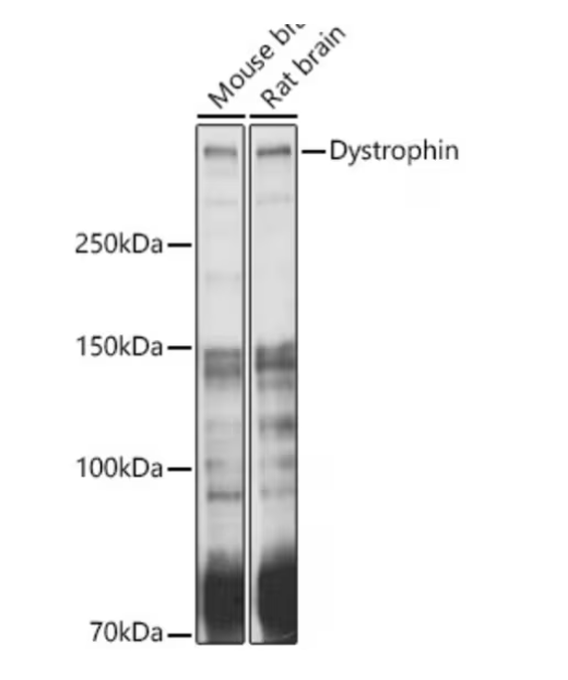
Dystrophin, a crucial protein for maintaining muscle integrity, is severely deficient in Duchenne Muscular Dystrophy (DMD) patients. Quantifying dystrophin restoration following therapeutic interventions is essential for evaluating treatment efficacy. Western blotting, a widely used protein analysis technique, plays a pivotal role in this quantification, with anti-dystrophin antibodies being central to its success.
Importance of Anti-Dystrophin Antibodies
Anti-dystrophin antibodies are specifically designed to detect and bind dystrophin protein, making them indispensable for Western blot analysis in DMD research. These antibodies enable:
- ✅ Detection of Dystrophin Rescue: By targeting dystrophin, these antibodies help visualize protein presence in treated versus untreated samples, indicating the extent of therapeutic rescue.
- ✅ Quantification: Signal intensity correlates with protein abundance, allowing researchers to quantify dystrophin levels compared to control samples.
- ✅ Therapeutic Validation: Essential for assessing exon-skipping therapies, gene therapies, and other dystrophin-restoring strategies.
Western Blot Methodology
A comprehensive methodology for Western blotting using anti-dystrophin antibodies emphasizes key considerations such as:
- Antibody Specificity: Ensuring antibodies recognize dystrophin without cross-reactivity.
- Sensitivity: Selecting high-affinity antibodies that detect even low dystrophin levels.
- Proper Controls: Including healthy and DMD samples for accurate comparisons.
Conclusion
By standardizing the use of well-validated anti-dystrophin antibodies, researchers can ensure consistent and reproducible results, driving progress toward effective DMD treatments.